
Macrophage barrier controls inflammation
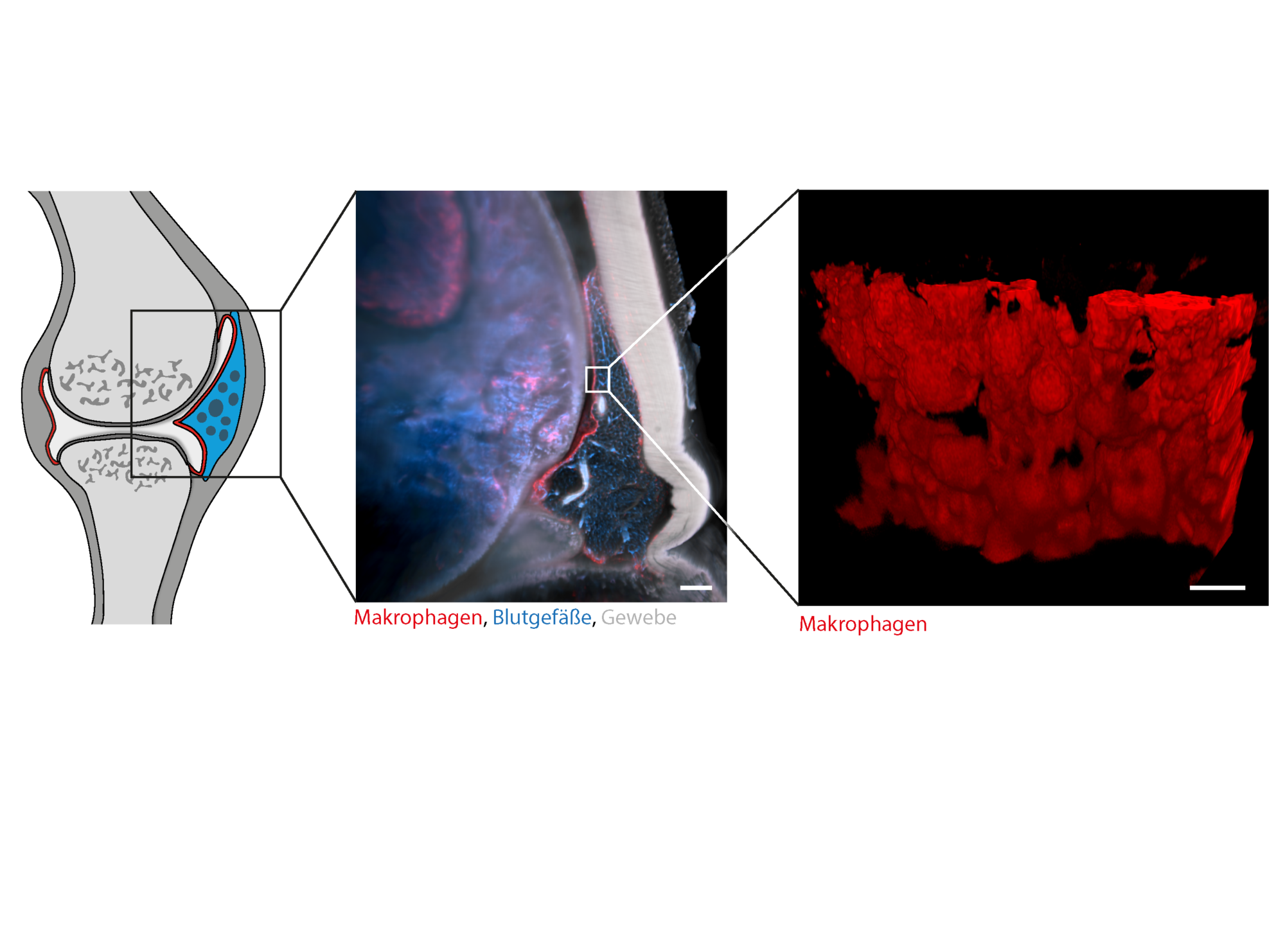
The exact causes of chronic inflammatory joint diseases such as rheumatoid arthritis are still poorly understood. A team of researchers around Prof. Gerhard Krönke has now chosen a new approach to better understand the underlying mechanisms. Stephan Culemann and Dr. Anika Grüneboom (Internal Medicine 3 – Rheumatology and Immunology) succeeded for the first time in making inflamed joints transparent and thus transparent to light with the aid of a specially developed technique. Subsequently, they examined these using three-dimensional molecular imaging. They gained surprising insights into how our immune system works. Among other things, it was shown that healthy joints are surrounded by a constantly self-renewing membrane of anti-inflammatory immune cells (macrophages). While this barrier of macrophages usually protects our joints from possible attacks of our own immune system, rheumatoid arthritis leads to a failure of this protective mechanism and thus to a sudden immigration of incorrectly activated immune cells, which ultimately cause joint inflammation and destruction. They have recently published their results in the magazine “Nature “.